AGE RELATED MACULAR DEGENERATION
Age related macular degeneration or AMD is an eye condition that occurs with aging. There are usually no obvious causes for this condition. However AMD affects the macula severely after the age of 50 leading to vision loss.
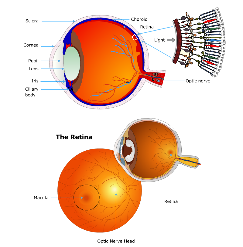
The retina and the macula
The retina is a curtain of cells that line the back of the eye. It is made up of special light sensitive cells called rods and cones. Light enters the eyes and passes through the lens which focuses the light on to the retina. The light causes electrochemical changes in the retinal cells leading to stimulation of the nerve cells supplying the retina. These nerve cells via the optic nerve carry the visual information to the brain that allows us to see an image. The retina contains the retinal pigment epithelium (RPE), the rods and cone cells and an inner lining Bruch's membrane.
The macula is the small part or spot that lies near the centre of the retina. The macula is where the vision is focused and is the best point of vision. The central and most important part of the macula is called the fovea. The outer part of the retina is responsible for peripheral vision.
AMD is one of the commonest forms of irreversible visual loss in patients over 50 years of age worldwide. 10% of 65-75 year-olds and 30% of over 75 year-olds are found to be affected to a certain degree with this condition. 1.7% of the population over 50 years of age in addition has severe disease and 18% of population over 85 have severe AMD. With rise of aging population, AMD incidence is on the rise.
Risk factors that increase the chances for AMD include :
- Increasing age Smoking is one of the well know risk factors for AMD
- There is some evidence that presence of hypermetropia or far sightedness may be a risk factor for AMD.
- Those with a previous cataract surgery may also be at a risk of AMD.
- Patients who have high blood pressure may also be at a high risk of developing AMD. Diabetes however is not a risk factor for AMD.
- Genes may play a role in risk of AMD. Some genes that run in families may be responsible for increased risk of AMD.
Similar to genetic link, ethnicity, gender and race are also associated with risk of AMD. White and Chinese people are more commonly affected than others while blacks are least affected. Women are said to be more affected in over 75 year-olds.
Pathology of AMD
In AMD the macula of the retina is affected. There is deposition of small collections of chemicals also called colloid bodies and commonly known as drusen. The drusen is deposited in the region between the retinal pigment epithelium (RPE) and the underlying Bruch's membrane. The changes are uncommon before the age of 45 years and as the persons ages the number and size of these drusen rises. In early stages these changes in the retina are called age-related maculopathy (ARM). After the changes reach a critical size in the macula, they are termed as age-related macular degeneration (AMD).
Classification of AMD
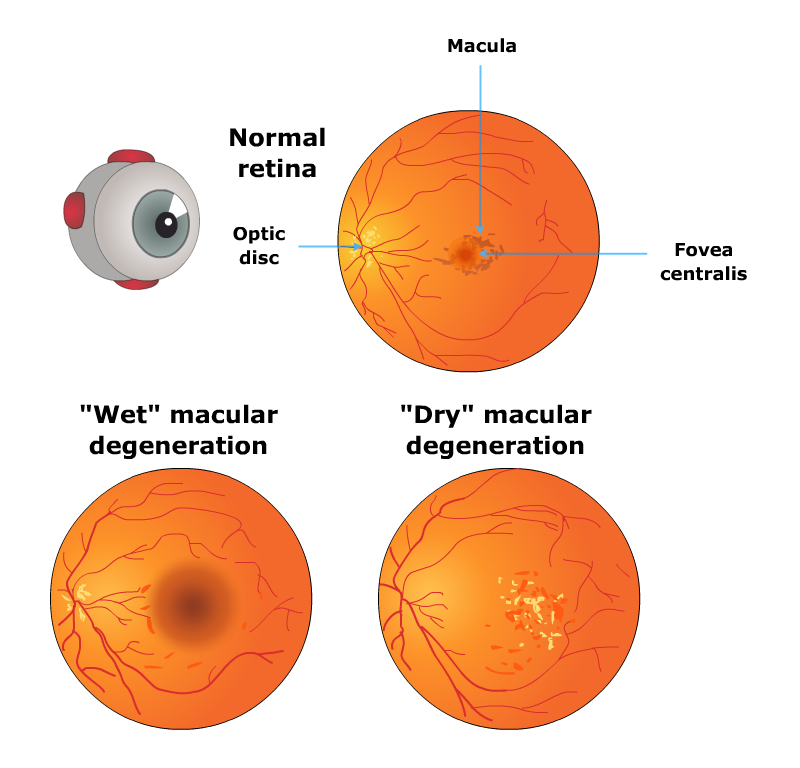
AMD can be classified in numerous manners. In Early AMD vision is preserved. There is early evidence of macular damage. The drusen appear small and there is an absence of pigmentary changes. These have a low risk of progression to large drusen to the tune of 0.4-3.0% over 5 years. Presence of large drusen however is associated with pigmentary changes progress to vision loss in almost 50% of cases. This often affects both eyes and second eye is affected in 60% cases within 5 years of affecting the first eye.
Types of AMD include
- Atrophic AMD – This is also called dry, non-exudative or geographic atrophy. Here the presence of drusen leads to progressive atrophy of the RPE, rods and cone cells and the choriocapillaris or the tiny blood vessels under the RPE. This is the commonest form of AMD occurring in 90% of cases
- Exudative AMD - This is also called wet or neovascular AMD. This is seen in 105 cases of AMD. In these cases there is an abnormal growth of choroidal blood vessels into the retina. This leads to formation of a vascular or blood filled membrane. This is also called sub-retinal neovascularisation (SRNV). In this there is accumulation of serous fluid within these membranes and this leads to detachment of the retina and loss of vision. There is formation of abnormal blood vessels as well. This leads to formation of a scar over the macula and loss of vision.
Age-related macular degeneration (AMD) may present with several initial symptoms that lead to progressive vision loss. Some of these symptoms include
- Difficulty in tasks that require visual focus like reading, driving and recognising faces
- There may be flashing lights. This is called photopsia or flickering or flashing lights
- Night glares
- Presence of floaters in the visual field
- visual hallucinations may be present. This is also called Charles Bonnet syndrome
- abnormal and reduced night vision
- In atrophic or dry AMD things may appear smaller or bigger than they are. This is called micropsia or macropsia respectively.
- In wet AMD there is profound central vision loss that may be sudden in case of bleeding
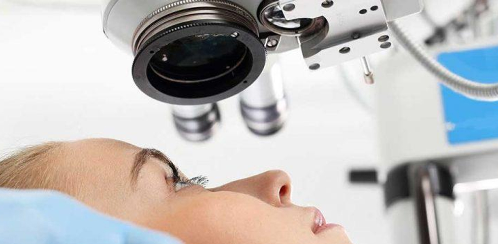
Your doctor may diagnose your condition by reviewing your medical and family history and conducting a complete eye exam. He or she may also do several other tests, including:
Examination of the back of your eye. Your eye doctor will put drops in your eyes to dilate them and use a special instrument to examine the back of your eye. He or she will look for a mottled appearance that's caused by drusen — yellow deposits that form under the retina. People with macular degeneration often have many drusen.
Test for defects in the center of your vision. During an eye examination, your eye doctor may use an Amsler grid to test for defects in the center of your vision. Macular degeneration may cause some of the straight lines in the grid to look faded, broken or distorted.
Fluorescein angiography. During this test, your doctor injects a colored dye into a vein in your arm. The dye travels to and highlights the blood vessels in your eye. A special camera takes several pictures as the dye travels through the blood vessels. The images will show if you have abnormal blood vessel or retinal changes.
Indocyanine green angiography. Like fluorescein angiography, this test uses an injected dye. It may be used to confirm the findings of a fluorescein angiography or to identify specific types of macular degeneration.
Optical coherence tomography. This noninvasive imaging test displays detailed cross-sectional images of the retina. It identifies areas of retina thinning, thickening or swelling. These can be caused by fluid accumulations from leaking blood vessels in and under your retina.
Being told you have age-related macular degeneration (AMD) can be frustrating and upsetting, as simple everyday tasks such as reading become more difficult.
It's estimated around a third of people with AMD may have some form of depression or anxiety.